Fibula. Part 3
Description
This section is from the book "The Anatomy Of The Human Skeleton", by J. Ernest Frazer. Also available from Amazon: The anatomy of the human skeleton.
Fibula. Part 3
Speaking generally, the articular surface on the femur (for the tibia) shows a decreasing curve as it is followed forward : that is, there is a segment of a small circle on the back of each condyle, the segment of a larger circle becomes apparent below, and a still larger one as the surface reaches the trochlear cartilage.
* In cases of genu recurvatum the ligament is stretched or wanting.
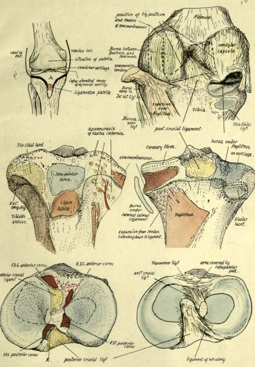
Fig. 131.-Right knee and upper end of tibia. Top left figure is a diagram of the synovial sac from the front, showing the trochlear part of the sac continued below each condyle into the condylar recesses, which are partly subdivided by the projection into them of the wedge-like semilunar cartilages. The synovial sac does not come down lower than the cleft between femur and tibia, whereas the " capsule," shown by the black lines, is brought down on the tibia in front to take in the tubercle and to be attached along the ligamentum patellae. Thus the area A is included on the front of the bone between the sac and the capsule, behind the patellar tendon, the interval being rilled by the infrapatellar pad and bursa (see Fig. 127). The front and side parts of the capsule are made by the aponeuroses of Vasti and Crureus, extending on each side to the lateral ligaments, and the deep fascia is blended with their superficial surfaces. Thus on the tibia, in the figure next below, the capsular attachment is represented by the Vasti, the lig. patella?, and the ilio-tibial band, while the infrapatellar bursa lies in position deep to these. On the right, the back of the joint is seen ; the tendon of Semimembranosus has been cut and turned back and its expansion (see Fig. 120) removed to show the crucial ligaments which occupy the notch between the condylar recesses and capsules ; the position of the tendon and expansion are indicated by interrupted lines. The opening for Popliteus exposes the outer meniscus, and the bursa round the tendon is carried on a cartilage-covered area on the tibia ; the muscle itself is not shown, but the expansion over it from Semimembranosus is seen. Below this the areas occupied by these several structures are shown on the tibia ; notice that, internal to insertion of Popliteus, fibres of the expansion from Semimembranosus reach the bone and roughen it, the upper ones passing under the ligament and the lower ones joining it. The capsule round the fibular facet is weak above, where the bursa may perforate it. The epiphysial line is shown as a thin green line in the middle figures. The lowest figures show the upper tibial surface. On the right the crucial ligs. and semilunar cartilages are in situ, cut away from the femur. Each cartilage lies on the tibia, the infrachondral part of the cavity being between them and the bone ; their thick margins are attached by coronary fibres to the bone, as shown in the middle figures, such fibres being, of course, blended with those of the condylar capsule behind. The free edges of the menisci rest on the tibia, their position being indicated by the interrupted lines in the left-hand figure, in which their cornual attachments and those of the crucial ligs. are also shown. ISC, ESC, inner and outer semilunar cartilages. At A" a part of the supra-chondral cavity reaches the bone between the post, crucial and internal posterior cornu.
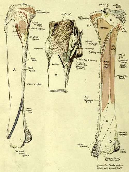
Fig. 134.-Right tibia, inner and posterior views. A, in the left-hand figure, is the subcutaneous inner surface of the shaft, crossed by the long saphenous vein. This surface is thus smooth, and slightly convex from side to side in conformity with the shape of the leg ; rather concave from above down in its lower half, owing to the increasing prominence of the lower end. In its upper part, however, the inner aspect of the bone shows secondary markings for Vastus internus. Gracilis (G), Sartorius (5), and Semitendinosus (5-7"). These are all deep to the deep fascia, which must therefore be carried in by them across the surface. Vastus internus is deep to the other three tendons, wherefore these, coming from behind and above, are inserted below and in front of the Vastus ; see the middle figure, where they are represented as a single insertion by a thick black line, and their situation over the Vastus and internal ligament is shown by interrupted lines. Vastus internus has a strong band that makes a definite rounded marking on the bone, the rest of its insertion being by more scattered fibres reaching almost to the tubercle (see Fig. 131), among which the internal inferior artery turns upwards. Internal lateral ligament is on the junction of inner and back surfaces, not very far above the inner end of the oblique line. B, area covered by Popliteus but not affording insertion to it ; at the extreme inner end of the triangular " popliteal surface " the bone is roughened by fibres, N, of Semimembranosus (expansion) reaching the bone as far down as the ligament. Below the oblique line the Tibialis posticus and Flexor longus digitorum mould the bone into surfaces in different planes, separated by the vertical line ; both arise by muscle fibre and hence show no secondary markings in their areas as a rule, but tendinous fibres in the upper part of Tib. post, may cause roughnesses near the upper part of the oblique line (see Fig. 133A). These deep flexors are covered in by the deep transverse fascia (fine green line). Tibialis posticus is directed downwards and inwards from its origin, therefore is in relation with the area C, while Flex. long, hallucis comes downwards and inwards from the fibula and lies on the area D ; between these the main vessels and nerve and tendon of F. long. dig. come into relation with the bone.
The internal lateral ligament is a long and strong band from the inner condyle (Fig. 122) to the tibia not very far above the level of the inner end of the oblique line (Fig. 134). Its upper attachment is about the centre of one part of the curve in which the tibia moves in flexion,as shown in Fig. 122,and overthis portion the tibia is efficiently held by the ligament : behind this the tension of the posterior crucial ligament holds the bones in apposition on the smaller curve. Observe that the internal lateral ligament comes down far below the proper level of (coronary) capsular attachment (this is indicated in Fig. 131), and below this the ligament covers the insertion of Semimembranosus, with a bursa over it, and is joined lower down by some fibres of the lower expansion from this tendon. Here it also crosses the lower articular vessels. Where it lies on the coronary fibres the ligament is attached to them, and through them has some connection with the margin of the internal semilunar cartilage.
Continue to:
- prev: Fibula. Part 2
- Table of Contents
- next: Fibula. Part 4