Development Of Ossification
Description
This section is from the book "The Anatomy Of The Human Skeleton", by J. Ernest Frazer. Also available from Amazon: The anatomy of the human skeleton.
Development Of Ossification
The innominate bone is represented in the early embryonic stages by a mass of mesenchyme which is situated in the base of the limb bud : about the beginning of the second month chondrification commences in this, the mesenchyme comes into relation with the vertebral region in the sixth week, and the cartilage is in position in the seventh week. There are three centres of chondrification corresponding with the three primary parts of the bone, and these fuse and form a shallow acetabulum in the seventh week. By the end of the second month the two innominate plates meet at the symphysis, and the pubic chondrification extends towards this later.
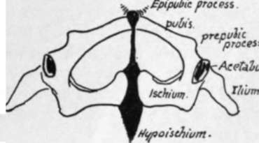
Fig. 113.-Pelvis of lizard seen from below. Cartilage black.
Ossification commences now, that is at the end of the eighth week, as the first of the primary centres, that for the Ilium. The centre for the Ischium is found just before the fourth month, and that for the pubis about a month later. These primary centres appear in their regions near the acetabulum.
At birth each of these main centres has formed a small piece of the corresponding acetabular wall, the rest of the hollow being cartilaginous, as are also the iliac crest, the front margin of the great sciatic notch, the region of the ilio-pectineal eminence, the region of the symphysis, and the ischio-pubic ramus.
Ossification extends slowly in this cartilage, so that about the tenth or eleventh year the ischial and pubic bones have met in the lower ramus, and just after this acetabular centres appear in the triradiate cartilage, which still separates the three primary bones as they have slowly extended on to the floor of the acetabulum.
The acetabular centres appear to be very variable in their number : they may join the neighbouring bones, or may fuse to form a single separate ossicle, the os acetabuli, which ultimately fuses with the pubic mass. In this way the acetabular region is consolidated by the age of puberty, fusion occurring first on the pelvic aspect ; but the solid bone is still edged by cartilage on the crest, along the front and back borders in places, in the region of the symphysis, and along the back part of the margin of the lower ramus.
In this cartilaginous border secondary centres appear as soon as the bone is consolidated, at or just after puberty. They are :-
(1) For the anterior superior spine and front part of crest ;
(2) For posterior superior spine and back part of crest ;
(3) For anterior inferior spine ;
(4) For spine of Ischium ;
(5) For marginal surface of ischial tuberosity (hypoischium) ;
(6) Somewhat later, for angle of symphysis ; and
(7) For spine, apparently not constant.
All these secondary centres are usually fused with the main mass by the age of twenty or twenty-one, the last two being a few years later in consolidating.
Continue to:
- prev: Borders. Continued
- Table of Contents
- next: Femur