Detailed Consideration Of Os Innominatum
Description
This section is from the book "The Anatomy Of The Human Skeleton", by J. Ernest Frazer. Also available from Amazon: The anatomy of the human skeleton.
Detailed Consideration Of Os Innominatum
The general " build " of the bone is associated with the transference of weight trom the vertebral column to the heads of the femora. The bone can be placed in the position it occupies in the complete pelvis by holding it so that the plane of its sym-physial articular surface is sagittal, while the cotyloid notch is opening directly downwards : when held thus it can be seen that the os innominatum admits of division into two parts that he in different planes. The upper and back portion looks in general backward and outward, whereas the lower and front parts looks forward and outward. The two planes cut each other nearly at a right angle, and the upper one roughly corresponds with the Ilium, the lower with the Ischium and pubis.
The scheme in Fig. 103 illustrates this : a is the upper and back plane and b the lower and front one. The projection forwards of a could be taken to Represent the schematic position of the anterior superior spine, while the hinder end of b would represent the projection of the tuberosity of the Ischium. The transition from one plane to the other is comparatively abrupt on the outer side, and occurs about a line drawn from below the Ischial spine to the anterior superior spine : this places the acetabulum on the lower segment, but, as can be seen on the other side of the scheme and is apparent on the bone, the part of the wall of this cavity that rests on the head of the femur and transmits weight to it is supported by the front edge of the upper plane, which carries the weight to it from the sacrum. Accordingly we find that that portion of the Ilium between the auricular surface and the top of the acetabulum is very thick and strong, and forms the acetabular rim along the line of transition from one plane to the other.
On the inner side, however, the transition is modified by the curved bar c of the ilio-pectineal line, whose mechanical function is concerned with the mode of transmission of the weight from the sacrum to the ilium.
The appearance of a section through the pelvis along the ilio-pectineal line would be in its essentials like the scheme in the figure : notice from the direction of the articular surfaces that the sacrum is not wedged down between the two hip bones, thus tending to drive them apart, but is suspended from them by posterior sacro-iliac ligaments (*), and therefore would draw them together as it throws weight on them. The bracket c counteracts the inward bending of a on b and makes the whole structure strong and rigid, dispensing with the necessity for strong anterior sacro-iliac ligaments to resist the separation of sacrum and ilium in front.
In this way we see why there must be such a large ligamentous area above and behind the auricular surface, separating it from the post-vertebral muscle area, with a very ill-marked ligamentous line below and in front of it, and at the same time it becomes clear that the pubic symphysis is not concerned in carrying or transmitting the weight of the trunk, and is on this account not provided with excessively strong ligaments.
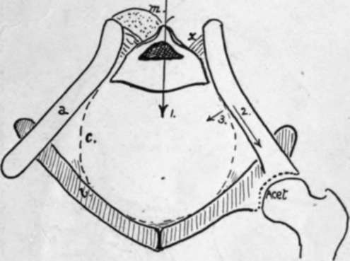
Fig. 103.-Scheme to show the mechanical build of the pelvis ; it practically represents a frontal section. Explanation in text.
Continue to:
- prev: Pelvis
- Table of Contents
- next: The Acetabular Region