Costal Cartilages. Part 5
Description
This section is from the book "The Anatomy Of The Human Skeleton", by J. Ernest Frazer. Also available from Amazon: The anatomy of the human skeleton.
Costal Cartilages. Part 5
The secondary markings made by the outer column of the Erector spinae and the aponeurosis can be seen on all the bones except the first : even on the last rib, although there is no true angle in the form of a bend of the shaft, yet there is frequently a secondary marking in series with that on the eleventh.
There is another thing to be noticed in connection with the angle: the pleural surface of the rib at this part of the bone shows a tendency to narrow somewhat in the upper ribs, and this reaches such a degree in the second that the pleural surface for a little distance is nearly linear on this bone, widening on the neck and on the under surface of the shaft. The meaning is not clear, but the result appears to be attained by the encroachment of the internal intercostal above, and this muscle with the subcostal groove below, and it is one of the minor points which, taken together, can be used to distinguish upper from lower ribs. In connection with the hinder parts of the ribs it remains to point out that the upward curve formed by these with their shafts is most marked in the seventh rib and decreases from this. This is best shown by putting the bones in a row, resting each bone on the lower border of the shaft as far as possible, when the vertebral ends will be seen to be raised from the table and a hne joining them forms a curve with its highest point made by the seventh head and the lowest by the second and tenth.
There is not much to be added to what has already been said about the shafts beyond the angles (pp. 48-50).
There is a tendency to the formation of a supra-costal groove between the attachments of the intercostal muscles on the upper ribs. The subcostal groove is perhaps better marked in the lower ribs-except 10, II, and 12-than in the higher : the floor of the groove is pierced by several small foramina directed towards the vertebral end of the bone. The lower margin of the shaft is sometimes drawn down to form a prominent flange, at and outside the angle, especially in the eighth and ninth ribs, a condition well marked in the foetus : it is interesting to note that downgrowths from the lower margins of the middle ribs, forming uncinate processes, are found among the Sauropsida, in crocodiles, some lizards, and many birds.
The general relations of the upper surface of the first rib have been already pointed out (p. 50). The extent of insertion of Scalenus medius is shown in Fig. 43. There is no distinct Levator for this rib, the muscle being continuous with the mass of Scalene fibres, but the area that receives what are apparently corresponding fibres is indicated in the figure : it is seen to extend in beyond the level of the tubercle.
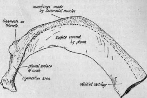
Fig. 44.-Lower surface of first rib.
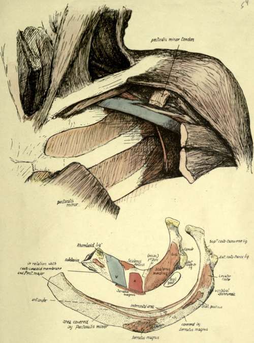
43--The lower figures are the first two ribs seen from above. The upper figure is from a dissection to show the course of the contents of the axillary sheath. Observe that these go backwards as well as outwards from the first rib, so that they come to lie on the Serratus magnus at once, even though this muscle arises comparatively far back on the two upper ribs. This haves the front portions of the ribs uncovered by either the muscle or the vessels, and here the pectoral structures must come into relation with the ribs : the first rib is here in relation with the costo-coracoid membrane and the second rib with I Vctoralis minor. Areas for both these structures may be seen on the second rib, as is shown in the lowest figure, and the anterior angle marks the continuity of the two areas. The lower figures require little comment, but it may be pointed out that the Subclavian and rhomboid attachments on the first rib appear there largely because there is some calcification of the cartilage, that the sharp groove for the ist dorsal nerve is not always evident, and the posterior part of Scalenus medium insertion, marked off by a dotted line and labelled LC, probably represents the Levator costa? found as a separate muscle lower down.
Fig. 45.-Last rib. Right side. See text.
Fig. 46.-Deep view of front wall. T, Triangularis sierni.
Its inner margin affords attachment to Sibson's fascia (and the Scalenus pleuralis if present), and its outer margin has a marking about half-way round for the origin of Serratus magnus : behind this the Scalenus posticus crosses the margin, just in front of the tubercle, and passes deep to the digitation of the Serratus to reach the second rib. In front of the origin of Serratus magnus the margin is in relation to the costo-coracoid membrane and Pectoralis major.
The lower surface of the first rib is nearly altogether pleural in its front half, the intercostal muscles being only attached along the outer margin. The various areas are shown in Fig. 44.
The narrowing of the pleural surface to a hne opposite and beyond the angle in the second rib has been noticed, as has also the smallness of the insertion of Levator costae and its continuity with the upper external intercostal. Pass the finger along the most prominent convexity of the bone just outside the angle and an irregularity will be detected which marks the attachment of Serratus posticus superior to the rib. Above and in front of this, near the upper margin, is another impression for Scalenus posticus, and these two muscles lie deep to Serratus magnus. The origin of Serratus magnus is from the prominent boss on the shaft, and immediately in front of this is a flatness, or even a shallow concavity, about an inch and a half broad, on which the Pectorahs minor rests when the arm is hanging by the side. These relations are shown in Fig. 43.
Continue to: