The Central Artery
Description
This section is from the book "Anatomy Of The Arteries Of The Human Body", by John Hatch Power. Also available from Amazon: Anatomy of the Arteries of the Human Body, with the Descriptive Anatomy of the Heart.
The Central Artery
The Central Artery of the Retina is extremely minute; it arises at the outer side of the optic nerve, pierces its coats, and runs forwards through its centre to arrive at the retina, on the internal surface of which it forms a vascular expansion which may be traced as far forwards as the ciliary processes. Immediately on escaping from the optic nerve, it gives off a branch, the Artery of Zinn, which runs from behind forwards through the centre of the vitreous humor, and contained within a sheath formed by the hyaloid membrane, called the hyaloid canal: it sends numerous small branches to the hyaloid membrane: in front it ramifies on the posterior part of the capsule of the lens, and in the foetus its branches have been traced to the membrana pupillaris. This artery occasionally arises from one of the ciliary arteries.
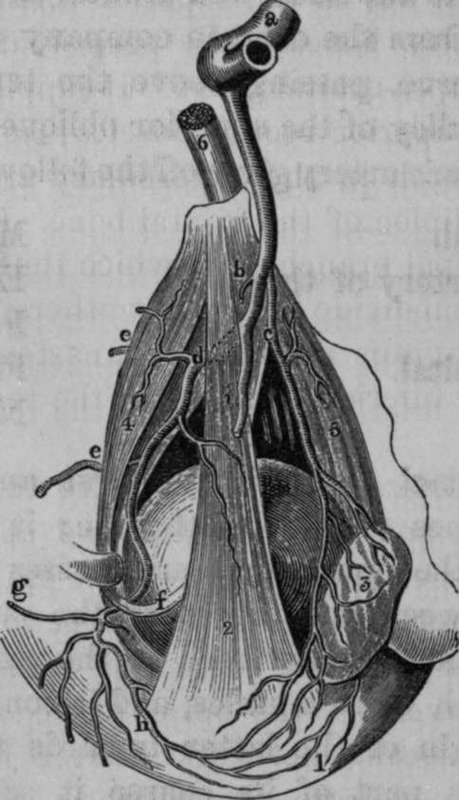
Fig. 17. Dissection of some of the branches of the Ophthalmic Artery.
1, Anastomosis between the Lachrymal and Superior Palpebral Arteries. 2, Levator Palpebrae Superioris Muscle. 3, The Lachrymal Gland. 4, Superior Oblique Muscle. 5, External Rectus Muscle. 6, Optic Nerve, a, Last turn of Internal Carotid Artery from which is given off the Ophthalmic Artery, c, Lachrymal Artery, d, Trunk of Ophthalmic Artery after having passed beneath the Levator Palpebrae and Superior Rectus Muscles, e, e, Anterior and Posterior Ethmoidal Arteries, f. Tendon of Superior Oblique Muscle after having passed through its pulley, g, Nasal Artery, h, Small portion of Superior Rectus Muscle, i, Supra-Orbital Artery cut across.
Continue to: