The Artery Of The Corpus Cavernosum
Description
This section is from the book "Anatomy Of The Arteries Of The Human Body", by John Hatch Power. Also available from Amazon: Anatomy of the Arteries of the Human Body, with the Descriptive Anatomy of the Heart.
The Artery Of The Corpus Cavernosum
This artery arises from the pudic immediately after it has passed through the anterior layer of the triangular ligament: it then pierces the crus penis, and advances through the corpus cavernosum, distributing its branches on either side, and gradually approaching the middle line. It communicates through the septum pectiniforme with the artery of the opposite side, and ramifies in the areolar tissue of the corpus cavernosum.
The following is Muller's opinion as to the distribution of the arteries of the corpus cavernosum :" The arteries of the corpus cavernosum have two sets of branches:the one set are the ultimate ramuscules, which terminate in the minute radicles of the veins, and are destined for the nutrition of the part; the other set come off from the side of the arteries, and consist of short, slightly curled branches, terminating abruptly by a rounded, apparently closed extremity, turned back somewhat on itself. These are sometimes single; sometimes several arise by one stem, forming a tuft. I have named them arteriae helicinae. They project into the venous cells, and are found principally in the posterior part of the corpora cavernosa, and of the corpus spongiosum urethrae. They are not distinct in man. Although no openings can be discovered in the coats of these free arterial excrescences, yet there is no doubt but that it is through them that the blood, which is ordinarily carried into the texture of the corpora cavernosa by the minute nutrient branches of the arteries, is, in the act of erection, poured directly into the venous cells and sinuses. When the arteria corporis cavernosi is injected with size and vermilion, the injected matter always fills the venous cells; and if it is afterwards washed from them, the arteriae helicinae will be seen injected. The means by which, during life, they are enabled to force blood into the cells must be the increased attraction exerted between their coats and the blood by the nervous influence transmitted to them by the spinal cord, in consequence of which attraction an increased quantity of blood goes to them. This throws new light, at the same time, upon the mutual action of the blood and smaller vessels in other parts, and upon the phenomenon of active turgescence, or turgor vitalis. The blood is returned from the corpora cavernosa partly by small veins, running, at the sides and on the surface of these bodies, into the vena dorsalis, partly by deeper veins, which issue from the corpora cavernosa at their root, and enter immediately the venous plexus, situated behind the symphysis pubis. The fact, then, that the vena dorsalis does not return the blood from the deep veins, shows that no pressure on the former vein alone can cause accumulation of blood in the penis."*
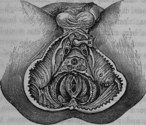
Fig. 54. Represents the Surgical Anatomy of the Male Perineum: the Crus Penis of each side divided and partly removed. The membranous portion of Urethra divided behind the Bulb; and the latter with the Corpus Spongiosum are turned forwards. The urethral opening in Triangular Ligament is seen, as well as the vessels between its layers. The anterior layer opened and some of it cut away.
B, The Bulb. C, Cowper's Glands receiving twigs from the Artery of the Bulb. D. D, The two Crura Penis. E, E, The Triangular Ligament or deep Perineal Fascia; a portion of its anterior layer removed. F, The Anus. G, G. The Tuberosities of the Ischia. H, The Coccyx. I, I, The Great Glutaei Muscles. K, K. The Levatores Ani Muscles partly removed. L, The Artery of the Bulb divided. M, The Urethral opening in Triangular Ligament. N, N, The Rectum. O, The Great Sciatic Ligament.
Continue to: