Operation Of Tying The Common Iliac Artery
Description
This section is from the book "Anatomy Of The Arteries Of The Human Body", by John Hatch Power. Also available from Amazon: Anatomy of the Arteries of the Human Body, with the Descriptive Anatomy of the Heart.
Operation Of Tying The Common Iliac Artery
The operation of tying the common iliac artery has been performed upwards of twenty times on the human subject; first, by Dr. Wm. Gibson, of Philadelphia, in 1812, in a case of gun-shot wound; the patient died from hemorrhage in thirteen days after the operations : it was tied in March, 1827, by Valentine Mott, of New York; and in the year following by Sir P. Crampton in this city. It has also been tied by Salamon, Liston, Guthrie, Syme, Deguise, Perigof, Post, Stevens, Peace, Stanley, Hey, and Lyon. Out of all these cases nearly two-thirds of them terminated successfully. Mott's case was successful ; and as it contains a great deal of important and interesting information, we shall detail it at length.
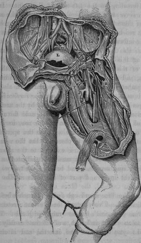
Fig. 43. Represents the Surgical Anatomy of the Iliac and Femoral Arteries.
A, Bifurcation of the Abdominal Aorta. B, The Anterior Superior Iliac Spine. C, Bifurcation of left common Iliac Artery. D. Poupart's Ligament. E. E*, The right and left Iliac Muscles, with the Inferior Musculo- or Inguino-Cutaneous Nerve of each side. F, The Inferior Vena Cava. O, Bifurcation of the right Common Iliac Artery. H, H*, The right and left Common Iliac Veins. I, I*, The right and left External Iliac Arteries, each crossed by the Circumflexa Ilii Vein. K, K*, The right and left External Iliac Veins. L, The Urinary Bladder, covered by Peritoneum. M, The Rectum, divided and tied. N, The Profunda Branch of the Femoral Artery. O, The Femoral Vein; o, the Saphena Vein. P, The Anterior Crural Nerve. Q, The Sartorius Muscle, cut. R, The Rectus Muscle. S, Pectineus Muscle. T, The Adductor Longus. U, The Gracilis Muscle. V, The Opening or Entrance into Hunter's Canal, with the strong Fibrous Structure given off by the Adductor Longus to the Vastus Interuus. g, g, The right and left Ureters.
Continue to: