General Description Of The Heart
Description
This section is from the book "Anatomy Of The Arteries Of The Human Body", by John Hatch Power. Also available from Amazon: Anatomy of the Arteries of the Human Body, with the Descriptive Anatomy of the Heart.
General Description Of The Heart
The Heart is a hollow muscular organ of a somewhat conical form, consisting of four chambers, grouped together so as to form an individual mass; two of these are called the auricles, the other two the ventricles. The apex of the heart is formed (in the adult) by the extremity of the left ventricle; and looks downwards, forwards, and to the left side, towards the interval between the fifth and sixth ribs: in many subjects it is curved a little backwards. The base is turned upwards, backwards, and to the right side, and corresponds to the right side of the fifth, sixth, seventh, and, sometimes, partly to the eighth, dorsal vertebrae. The posterior inferior surface is flat and triangular, and the anterior superior surface convex and more extensive : these surfaces are separated by two margins: the anterior margin is thin, and looks downwards, forwards, and to the right side: the posterior margin, which is shorter but considerably thicker, looks in the opposite direction.
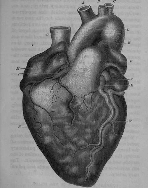
Fig. 1. Anterior View of the Heart.
A, Arteria Innominata. B, Left Carotid Artery. C, Left Subclavian Artery. D, Aorta. E, Remains of Ductus Arteriosus. F, Pulmonary Artery. G, Superior or Descending Vena Cava. H, Right Auricle. I, Posterior or Right Coronary Artery. K, Left Auricular Appendix. L, Anterior or Left Coronary Artery. M, Left Coronary Vein. N. Anterior surface of Right Ventricle.
The chief bulk of the heart is formed by the ventricles, particularly by the left; and the auricles seem like appendages situated at its base.
The two auricles are situated at the base of the ventricles, and towards its posterior part. When injected, and viewed as one, they form a crescentic mass, the concavity of which looks forwards and rather upwards, and embraces within it the aorta and pulmonary artery. The convexity looks backwards and somewhat downwards. The two extremities of the crescent are formed by the tips of the right and left auricular appendices.
The two ventricles taken together form a conical mass, which gives the peculiar form to the heart; it is obliquely situated, the apex being directed downwards, forwards, and to the left side; the base upwards, backwards, and to the right side.
The anterior superior surface of this mass is convex, and presents a fissure which runs from the base to the right side of the apex; this fissure lodges the anterior coronary artery and vein, and a quantity of fat, and divides the anterior surface into a right and left portion : the latter is formed by the anterior surface of the left ventricle, and the former, which is much larger, is formed by the anterior surface of the right ventricle. In this latter situation, Dr. Baillie has described a white opaque spot, like a thickening of the serous layer covering the heart: it is sometimes not broader than a sixpence; at other times broader than a crown piece; "it is so very common, that it can hardly be considered as a disease."*
The posterior inferior surface of the ventricular mass, which is less extensive than the superior, is nearly flat, and rests on the superior surface of the diaphragm, with the interposition of the base of the pericardium. This surface also is divided into two portions of unequal size by a fissure running from the base to the right side of the apex, and containing within it the posterior coronary artery and vein, and some fatty tissue: the larger portion is formed by the left ventricle, the remaining portion by the right.
The anterior margin of the ventricular mass is thin, longer than the posterior, and formed by the right ventricle : the posterior margin is thick and convex, and is partly lodged, with the intervention of the pericardium, in a depression of the left lung, and is formed by the left ventricle. The apex is formed, in the adult, entirely by the left ventricle; and the base presents for examination the following parts: anteriorly, a funnel-shaped projection of the right ventricle which passes upwards, and is termed the infundibulum, and from which arises the pulmonary artery: on a posterior plane, concealed by the infundibulum, and more to the right side than the orifice of the pulmonary artery, is the origin of the aorta from the base of the left ventricle. Behind these two orifices the base of the ventricular mass presents a circular fissure, circumscribing that portion of it which corresponds to the auricles: this fissure is very deep posteriorly: lastly, the base of the ventricular mass is cut obliquely downwards and backwards at the expense of the posterior inferior surface, which is consequently shorter than the anterior superior surface.
* Baillie's Morbid Anatomy, by Wardrop, p. 54.
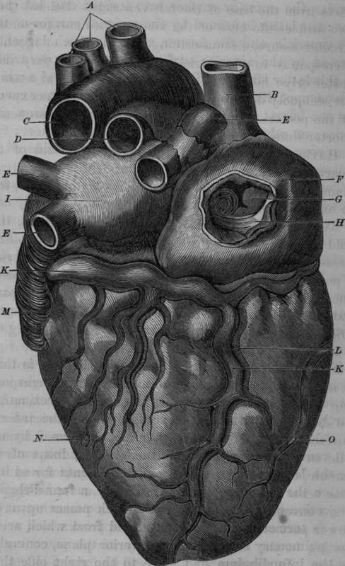
Fig. 2. Posterior View of the Heart.
A, Orifices of the Arteria Innominata, Left Carotid and Left Subclavian Arteries. B, Superior Vena Cava. C, Orifice of the Aorta. D. Orifice of the Pulmonary Artery. E, E, E, Orifices of the Pulmonary Veins. F, Right Auricle. G, Orifice of the Inferior Vena Cava. H, Eustachian Valve. I, Left Auricle. K, Posterior Coronary Vein. L, Posterior Coronary Artery. M, Left Auricular Appendix. N, Posterior part of Left Ventricle. O, Posterior part of Right Ventricle.
Having thus described the external surface of the heart, we may now proceed to consider individually its chambers, which are, as we have already observed, four in number: two auricles and two ventricles.
Continue to: