Tumors Of The Hair Follicles
Description
This section is from the book "Skin Cancer", by Henry H. Hazen, A.B., M.D.. Also available from Amazon: Skin Cancer.
Tumors Of The Hair Follicles
The hair follicle has different layers, just as has the skin, and epithelial growths may originate from any of these layers. Krompecher* states that he has been able to recognize two tumors of the face as springing from the basal layers of the hair follicles, and various other pathologists have reported similar observations. It is certain that, as our knowledge and technic improve, we shall later be able to identify neoplasms as springing from some of the numerous layers.
JUnna: Histopathology of Diseases of the Skin, New York, 1896. 2 Krompecher: Der Basalzellenkrebs.
At the present time .two different clinical and pathological varieties of tumor arising from the basal layer have been differentiated. The first is the multiple benign cystic epithelioma, subvariety of Von Zarisch, which is clinically indistinguishable from the more common type known as the Brooke-Fordyce* variety, where the growth is from the basal layers of the epithelium proper. These tumors will be fully discussed in the chapter dealing with the multiple benign tumors.
The second is the rodent ulcer type, or the trichoepithelioma of some authors.
The case reported by Bloodgood* is a fair illustration of this type. It occurred upon the cheek, just anterior to the angle of the jaw, in an elderly man, having been present for fourteen years as "a small elevated area covered with hair," probably a nevus of the hairy type. During the last year it had grown to about 7 or 8 cm. in diameter, and, as the result of the use of a caustic paste, there was a slight superficial ulceration, with considerable induration at the edge of the solid portion of the neoplasm. The tumor was freely movable over the cheek muscles. It was easily excised under cocaine, and it was then found that there was a zone of normal fat between the neoplasm and the underlying muscles. At the end of fourteen years there had been no recurrence. On section of the tumor the gross appearance showed large hair follicles, containing hairs in the depth of the tumor. Between the follicles the normal tissue was replaced by "a firm, white, granular tissue, divided into alveoli by a firm connective tissue stroma. Here and there were a few cavities, containing finely granular material-that is, the naked eye appearance of an epithelioma spinocellulare malignum." Histologically, however, the cells were not those of a prickle-celled carcinoma, but more nearly resembled basal or cuboidal cells, and in places could distinctly be seen springing from the hair follicles.
In Bloodgood's laboratory the author had the opportunity of studying another case in which the tumors were of much slower growth, and in which the cells were distinctly basal in character.
Recently the author has had a rather interesting case in his own practice. The patient was a man of 45, referred by Dr. Balloch, who had been struck upon the head two years previously, while on a fishing trip in the north woods. A few months later there developed a nodule, which had slowly increased in diameter. When seen, there was on the right anterior quadrant of the scalp a large growth that closely resembled a typical rolled-edge rodent ulcer. It was at least 12 cm. in diameter, the edge was very hard, and firmly attached to the skin, but not to the underlying bone. No ulceration had taken place. The hair was sparse, just as it was upon the remainder of his scalp. As operation would have been very difficult because of the size of the growth, it was determined to try the x-ray, and the results were very gratifying, for the growth promptly healed and for over two years there has been no recurrence. Histologically, the picture was very similar to Bloodgood's case (Fig. 36). There were very large hair follicles extending deep into the tissues; practically the whole field was filled with them, packed so closely together that there was no room for any other tissues. The hypertrophy was almost confined to the basal layers, and from some of them escaping strands of cells could readily be distinguished. The follicles contained no hairs.
*Sutton: Jour. Amer. Med. Assn., 1912, lviii, 333. ♦Bloodgood: Prog. Med., Dec., 1904.
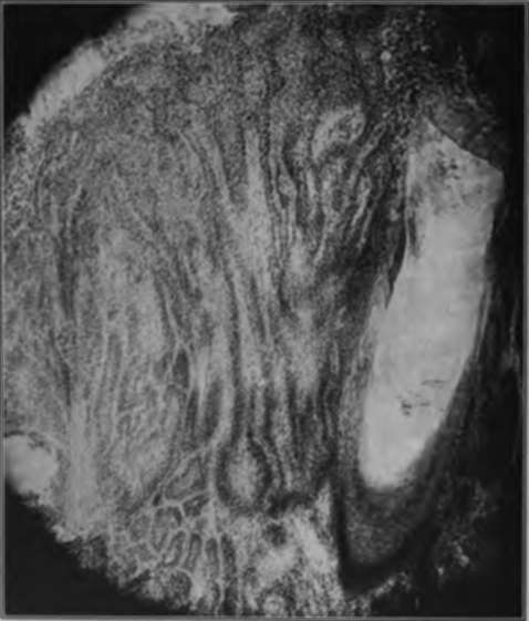
Fig. 36.-Carcinoma originating in hair follicles. Low-power photomicrograph. (Author's collection).
In addition to the various types of cases mentioned above, it must be borne in mind that some of the typical rodent ulcers, or basal-celled carcinomata, undoubtedly originate in the hair follicles.
Continue to:
- prev: Chapter VII. Benign And Malignant Tumors Of The Cutaneous Appendages
- Table of Contents
- next: Tumors Of The Sebaceous Glands
Tags
bookdome.com, books, online, free, old, antique, new, read, browse, download